Imaging in Oncology
(25) Rare Instance of Uterine Leiomyomatosis Vascular Extension Into the Cavoatrial Junction
Saturday, September 23, 2023
6:00 PM - 7:30 PM East Coast USA Time

Purpose: The purpose of this case study is to investigate the interesting complications that may arise from uterine Leiomyomas.
Uterine Leiomyomas are extremely common in the female population with a strong genetic role that plays part in the chance of development. The common complications of leiomyomas are: chronic pelvic pain, heavy menstrual bleeding, anemia, poor pregnancy outcomes, infertility.
In this case a 54 year old female patient presents to the emergency department with a chief complaint of heart palpitations of unknown chronicity. The patient has a history of 1x episode of idiopathic DVT anti coagulated with heparin in the past, history of stable, partially calcified uterine leiomyoma which has decreased in size 30% since menopause. Initial blood work does not show any abnormalities, d-dimer is not elevated. Patient’s vitals are within normal range at the time of exam.
Material and Methods: CT Abdomen/Pelvis was ordered to evaluate stability of known mass/bleed.
Subsequent MRI imagine was ordered to further evaluate initial findings.
Retrospective evaluation of previous out of network imaging for comparison to current imaging was performed.
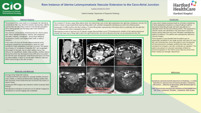
Results: The contrast CT shows a large filling defect within the internal iliac vein on the right extending to the IVC where it shows a large dependently layering filling defect with calcified components extending up to the cavoatrial junction.
Subsequent T2 MRI confirms that this is likely a T2 hypointense filling defect representing which is extending into the IVC.
Retrospective review of previous out of network images demonstrated a prior CTA performed for stability of the uterine leiomyoma showed that there was a filling defect within the right internal iliac vein confluence that had not yet extended into the IVC.
Conclusions: A case which initially presented itself as a female complaining of heart palpitations resulted in a unique finding of uterine leiomyoma extending through the local vasculature in a cephalad vector into the cavoatrial junction. This outgrowth of smooth muscle and calcification may have been a focus for ectopic pacing beats that may have ultimately manifested the patient’s symptoms.
The patient was subsequently referred to vascular surgery. Post evaluation it was decided that the patient was an appropriate candidate for two stage excision with open IVC and iliac vein thrombectomy.
